
Technology & PLAN
UNIQUE TECHNOLOGY FOR MEDICAL IMAGING
The new medical imaging method, Ultrasound Optical Tomography (UOT), combines ultrasound, laser spectroscopy and special quantum sensors to take high-resolution images of tissue oxygenation. Deficiencies in oxygenation can cause life-threatening acute conditions such as birth asphyxia in neonates, stroke and heart attack. The technology can give doctors the unique advantage of getting important information about the patient’s health more quickly and easily with good image resolution when rapid diagnosis is crucial, with a mobile device that can be placed in an emergency department or clinic.
Deviations in oxygenation are also characteristic of several forms of cancer tumors and can constitute a diagnostic marker. The functional imaging thus opens up the possibility of non-invasive or “digital” biopsy, i.e. providing functional information inside an organ, without physical sampling in, for example, breast cancer.
UNIQUE COMBINATION OF ULTRASOUND AND LASER SPECTROSCOPY
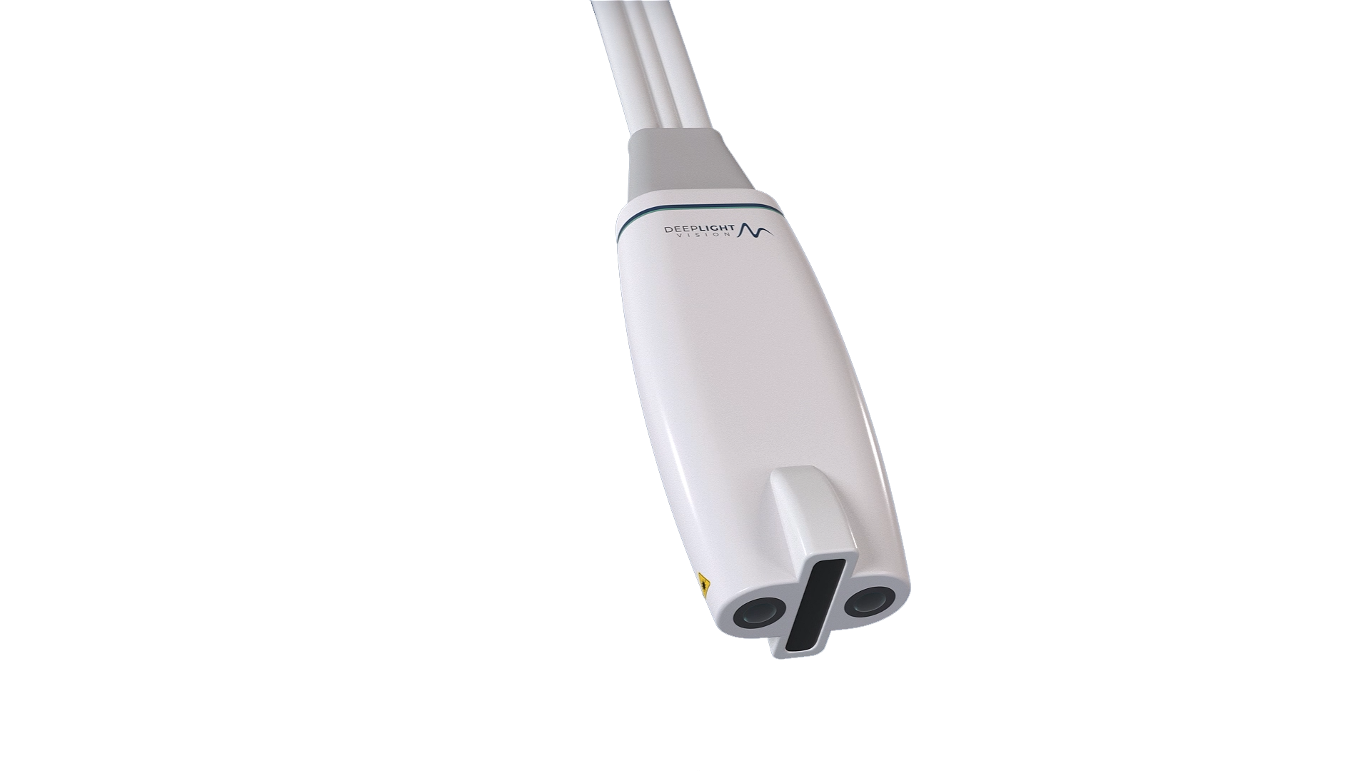
Deep Light Vision’s UOT technology integrates ultrasound, and laser spectroscopy. The UOT system combines ultrasound and laser spectroscopy. Laser spectroscopy uses light from a special type of laser to obtain detailed information about the oxygenation in the body. The laser spectroscopic part is related to the device, pulse oximeters, that doctors use to monitor blood oxygenation with a clamp around a finger. A pulse oximeter can measure the oxygenation in, for example, a finger, but does not provide image information. Deep Light Vision’s UOT system has the potential to provide a high-resolution image of the oxygenation deeper in the body in, for example, the brain or the heart.
WORLD LEADER IN QUANTUM SENSORS BASED ON "SLOW-LIGHT" FILTERS
Deep Light Vision and the research group in Lund are pioneers and world leaders in the development of so-called “slow-light” filters. The UOT technology uses special high-performance optical filters based on quantum technology developed in Lund. These “slow-light” filters have the property of sorting out, with extreme precision, the weak signal that occurs when laser light pulses sent into the body interact with the ultrasound field from an ultrasound scanner. This allows you to see detailed information about the oxygenation. Both the laser light pulses and the ultrasound field have low intensity and are harmless.
CLINICAL USE
When using the UOT system in healthcare, the system will have a handling similar to traditional ultrasound. An ultrasound probe combined with a laser transducer is passed over the tissue to be examined, and the image is presented in real time to the examining doctor or nurse.
DEVELOPMENT AND COMMERCIALIZATION PLAN
Our initial focus is on neonatal care and breast cancer diagnostics, with plans to expand into other patient groups subsequently. This strategy will establish clinical validity and address urgent medical needs.
For neonatal care, birth asphyxia is a leading cause of neonatal mortality and long-term neurological impairments, requiring fast and accurate early detection. The UOT system provides non-invasive, real-time oxygenation imaging, which has the potential to provide timely intervention in the critical hours after birth to prevent brain damage.
In breast cancer diagnostics, UOT would offer a non-invasive alternative for early detection, especially in challenging cases involving dense breast tissue or women under 50. UOT’s ability to provide real-time oxygenation data would allow clinicians to assess suspicious lesions more effectively and reduce unnecessary biopsies, enhancing the early detection of breast cancer.
By focusing on neonatal and breast cancer applications, UOT’s advanced diagnostic capabilities have the potential to significantly improve patient outcomes. Our phased development, coupled with expert partnerships, sets the stage for a successful market introduction.
1. UOT PROTOTYPE SYSTEM
A first UOT prototype system has been developed and tested in a lab environment with so-called tissue phantoms that mimic the properties of biological tissue. The tests have shown that the technology performs as planned.
2. CLINICAL RESEARCH STUDY
In the next step, a clinical research study is planned to be carried out with tests on humans. The purpose of the study is to investigate the system’s performance, which medical indication will be the company’s first priority, and to get feedback on the continued product development in order to subsequently develop a commercial system.
3. PRODUCT DEVELOPMENT
The further development is planned in stages in order to efficiently produce a product for medical technology market approval and later launch. Feedback from the clinical research study will provide direct input to the continued product development of the UOT system which will later be CE marked and commercialized.
4. CLINICAL STUDY FOR MARKET APPROVAL
When the product development is complete, clinical studies with the UOT system will be conducted to obtain market approval (CE marking in the EU and FDA approval in the USA).
BUSINESS MODEL
The offer to the market will include the UOT equipment, peripherals and service and support agreements. This can provide the company with recurring revenue streams during the product’s life cycle. Deep Light Vision will have the opportunity to bring the products to market ourselves, or seek cooperation with existing distributors or manufacturers of equipment for medical image diagnostics.